


FLOW VISUALIZATION TAKES CENTER STAGE AT TEDMED 2016 AS DR. PARTHO SENGUPTA, IN COLLABORATION WITH HITACHI, PRESENTS ADVANCED VISUALIZATION TECHNIQUES
Wallingford, CT, December 5, 2016 - Evaluation of cardiac hemodynamics progresses to a new level as presented by Dr. Partho Sengupta at TedMed 2016. In collaboration with Hitachi and others, Dr. Sengupta demonstrated a true ground-breaking technology; the evaluation of cardiac valves using holograms.
His presentation tells his personal story of an impressive journey through the diagnostic world of cardiac imaging that ultimately brought him to where he is today. His focus is strongly on discovering advanced visualization technologies to evaluate valvular form and function and how it is linked to the cardiac flow geometry. Some of Dr. Sengupta’s past work has been with PIV to demonstrate the valvular flow with vector information. He shared how he is now also using ultrasound to provide the vector information to evaluate this same flow geometry. He presents examples of PIV and Hitachi’s Vector Flow Mapping (VFM) analysis results side by side to illustrate the validity of ultrasound as a tool for vector mapping.
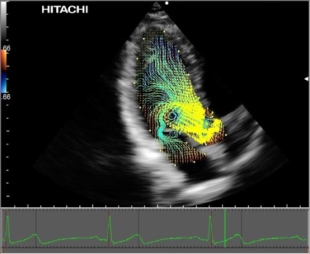
VFM using Velocity Vectors and Streamline display in a normal patient
As a look into the future, Dr. Sengupta then demonstrated how holograms will play a part in valvular and other cardiac diagnosis by showing an example of a mitral valve hologram. This example provided a remarkable snapshot into the future of evaluating cardiac physiological characteristics by using holograms.
Hitachi’s VFM stands in the forefront as an example of advanced hemodynamic analysis today. In the future, VFM along with hologram analysis are true leading-edge technologies that will certainly advance the diagnostics of cardiac health.